- BiologyDiscussion.com
- Follow Us On:
- Google Plus
- Publish Now


Essay on the Digestive System (For Students) | Human Physiology
ADVERTISEMENTS:
In this essay we will discuss about the digestive system in humans. After reading this essay you will learn about:- 1. Organs of Digestive System 2. Accessory Glands for Digestion of Foods.
Essay # 1. Organs of Digestive System:
Digestion means simplification of complex foods. It is the process of breaking various foodstuff into simple products. The complex foods like carbohydrates, proteins and fats are converted into glucose, amino acids and fatly acids respectively by the action of digestive enzymes. These simple substances enter into the blood circulation after absorption and then they are utilized by the body.
Digestive system consists of two main organs:
(1) Alimentary Canal
(2) Digestive Glands
1. Alimentary Canal:
This is also known as digestive tract or gastrointestinal tract. It is a long tube of varying diameter which begins at the mouth and ends at the anus. The length of this tube is about 8-9 meters. It opens at both the ends. The alimentary canal starts at the mouth into which cavity, the glands of the mouth pour the juice. As it passes backwards, it spreads into a funnel shaped cavity called-pharynx.
The tube then narrows into a soft muscular tube about ten inches in long, called the food pipe or gullet. This passes down the neck into the chest. It then opens into the stomach by piercing the diaphragm. The stomach is a large bag lying a little to the left under the diaphragm. It has two openings, one where the food pipe ends and the other where the intestines begin. The alimentary canal narrows again and passes into the small intestine which is about twenty two feet in length.
The first ten inches of the small intestine is called as Duodenum which forms a ‘C’ shaped loop. The rest of the small intestine is like a coiling tube, whose ends opens into a wide but comparatively short tube known as large intestine. It is about six feet long. The last part of the Large Intestine is known as Anus.
2. Digestive Glands:
Various digestive glands help in the digestion of foods.
(1) Salivary glands in the mouth,
(2) Gastric glands in the stomach
(3) Pancreas,
(5) Intestinal glands in small intestine.
All these digestive glands secrete digestive juices containing different enzymes which digest carbohydrate, protein and fatly foods.
Digestive juices:
Five digestive juices are secreted from digestive glands of the body. The enzymes present in these juices help in the digestion of different types of foods.
These juices are:
1. Salivary juice from salivary glands in mouth.
2. Gastric juice from Gastric glands in the stomach.
3. Pancreatic juice from Pancreas.
4. Intestinal juice from Small Intestine.
5. Bile juice from Liver.

Why so many digestive juices are necessary for digestion of food?
There are three reasons for the presence of so many digestive juices:
1. One digestive juice cannot digest three types of foods i.e. proteins, fats, and carbohydrates up to their completion.
2. One digestive juice cannot digest one particular type of food up to its completion, because food cannot remain in one place for a longer period of time.
3. The medium of action of enzymes present in different digestive juices are different. Some act on acidic medium and some on alkaline medium.
Digestion in Different Parts of Alimentary Canal:
The alimentary canal consists of the following organs in which foods are digested:
2. Oesophagus
4. Duodenum
5. Small Intestine
6. Large intestine
The mouth cavity is the front spread out end of the food pipe. The sides of the cavity are formed by the cheeks, the roof by the palate, and the floor by the tongue. When closed, it is bound in-front by the upper and the lower sets of teeth meeting in the middle. The opening at the back of the mouth is known as throat on each side of which there is a mass of tissue called tonsils. In the outside of the mouth cavity there is a slit like opening which is bounded by two soft movable lips.

You can Choose category
The Digestive Organ System
In people’s bodies, organs work together and form organ systems by combining with each other. One of the vital organ systems is the digestive one and consists of the gastrointestinal tract and organs such as the tongue, salivary glands, pancreas, liver, and gallbladder (Crash Course, 2015). This organ system is responsible for processing the food that is ingested in the mouth and is located mainly in the stomach.
Such a major organ system works with the help of positive and negative feedbacks caused by homeostasis, which preserves a certain pH level, the body temperature, and other measures in the state of balance (Amoeba Sisters, 2018). Some stimulus produces change to the body, which is detected by the receptor passing on the information within the input to the control center. Then, output carries back the received response, which is addressed to the stimulus in order to maintain homeostasis. The negative feedback to the stimulus reduces the effect caused by it, whereas the positive feedback increases it.
The positive feedback beneficial to the digestive system can be seen in bile acid production while eating fatty foods. In the balanced state of the organism, no or little bile acid is produced. However, appeared stimulus (meaning, fatty food) makes the liver produce the bile acid, which helps the digestive system process the food. I, similar to anyone else, experience this positive feedback during my lifetime, which helps me eat some unhealthy food sometimes and be sure that the digestive system will cope with it.
Concerning the example of the negative feedback that I ever had, I can remember the sweltering day of this summer, where the air conditioner helped my body to lower the temperature. In other words, the decreased temperature in the room became a stimulus to my skin receptors, which caused a lowering of the temperature of my body and made me feel more comfortable that day.
Crash Course. (2015). Introduction to anatomy and physiology: Crash course A&P #1 [Video]. YouTube. Web.
Amoeba Sisters (2018). Homeostasis and negative/positive feedback [Video]. YouTube. Web.

Essay on Digestive System
Students are often asked to write an essay on Digestive System in their schools and colleges. And if you’re also looking for the same, we have created 100-word, 250-word, and 500-word essays on the topic.
Let’s take a look…
100 Words Essay on Digestive System
Introduction to digestive system.
The digestive system is a group of organs that work together to change the food we eat into energy for our bodies. It’s like a food processing factory. It includes the mouth, esophagus, stomach, small intestine, large intestine, rectum, and anus.
Process of Digestion
Digestion starts in the mouth when we chew food. It then travels down the esophagus to the stomach. In the stomach, food is mixed with stomach acids to break it down into a liquid. This liquid then moves to the small intestine.
Role of Small Intestine
The small intestine plays a major role in digestion. Here, nutrients from the liquid food are absorbed into the bloodstream. The blood then carries these nutrients to all parts of the body. The leftover food, which the body can’t use, moves to the large intestine.
Role of Large Intestine
The large intestine is the last part of the digestive process. It absorbs water from the leftover food and turns it into waste. This waste then leaves the body through the rectum and anus. This whole process is known as digestion.
Also check:
- Paragraph on Digestive System
- Speech on Digestive System
250 Words Essay on Digestive System
What is the digestive system.
The digestive system is a group of organs that work together to change the food you eat into energy and basic nutrients to power your body. It is like a food processing plant that takes in raw materials (food) and turns them into something the body can use.
Parts of the Digestive System
The digestive system is made up of several parts. It starts with the mouth, where you chew and swallow your food. Then there’s the esophagus, a tube that carries food to your stomach. The stomach is like a mixer, churning and breaking down food into a liquid.
How Food Travels
From the stomach, the liquid food then goes into the small intestine. Here, it is broken down even more so your body can absorb the nutrients. Finally, what’s left goes into the large intestine, and then out of your body as waste.
The Role of the Liver and Pancreas
The liver and the pancreas also play important roles in digestion. The liver makes a juice called bile that helps to break down fats. The pancreas makes juices that help to break down carbohydrates, fats, and proteins.
Importance of the Digestive System
The digestive system is very important. Without it, our bodies wouldn’t get the nutrients they need. It keeps us healthy and gives us energy. So, remember to eat a balanced diet to keep your digestive system happy and healthy.
500 Words Essay on Digestive System
The digestive system: an introduction.
The digestive system is a group of organs that work together to change the food we eat into energy our bodies can use. It’s like a food processing factory inside our body. It includes the mouth, esophagus, stomach, small intestine, large intestine, rectum, and anus. The liver and pancreas also play a big role in digestion.
Starting Point: The Mouth
Digestion begins in the mouth. When we eat, our teeth break down the food into smaller pieces. Our saliva, a liquid made by the salivary glands, mixes with these pieces, making them easier to swallow. Saliva also starts the process of breaking down the food chemically.
The Esophagus: The Food Pipe
The esophagus is a long tube that connects the mouth to the stomach. It uses a process called peristalsis to move food. This process is like a wave of muscle contractions that pushes the food down into the stomach.
The Stomach: The Mixing Pot
The stomach is like a mixing pot. Here, the food is mixed with stomach acid and enzymes, which break it down into a liquid. This liquid is then sent to the small intestine.
The Small Intestine: The Nutrient Absorber
The small intestine is where most of the digestion happens. It is a long, coiled tube where nutrients from the food are absorbed into the bloodstream. The liver and pancreas help in this process. The liver makes bile, a substance that helps break down fats. The pancreas makes enzymes, which assist in breaking down proteins and carbohydrates.
The Large Intestine: The Water Saver
The large intestine, also known as the colon, is the final part of the digestive system. Its job is to absorb water from the remaining indigestible food matter, and then to pass useless waste material from the body.
The End of the Journey: The Rectum and Anus
The rectum and anus are the last parts of the digestive system. The rectum stores the waste until it’s ready to leave the body. Then, it passes through the anus and out of the body as feces.
Conclusion: The Importance of the Digestive System
The digestive system is vital for our survival. It turns the food we eat into nutrients that our body needs for energy, growth, and cell repair. Without it, we wouldn’t be able to live. So, next time when you are eating your favorite food, remember the amazing journey it takes through your body!
Remember, eating a balanced diet and drinking plenty of water can help keep your digestive system healthy and working well. Regular exercise is also important as it helps keep food moving through the digestive system, reducing the risk of constipation.
That’s it! I hope the essay helped you.
If you’re looking for more, here are essays on other interesting topics:
- Essay on Different Religions
- Essay on Diet And Nutrition
- Essay on Diet And Health
Apart from these, you can look at all the essays by clicking here .
Happy studying!
Leave a Reply Cancel reply
Your email address will not be published. Required fields are marked *
Save my name, email, and website in this browser for the next time I comment.

23.1 Overview of the Digestive System
Learning objectives.
By the end of this section, you will be able to:
- Describe the organs of the alimentary canal from proximal to distal, and briefly state their function
- Identify the accessory digestive organs and briefly state their function
- Describe the four fundamental tissue layers of the alimentary canal and the function of each layer
- Contrast the contributions of the enteric and autonomic nervous systems to digestive system functioning
- Explain how the peritoneum anchors the digestive organs
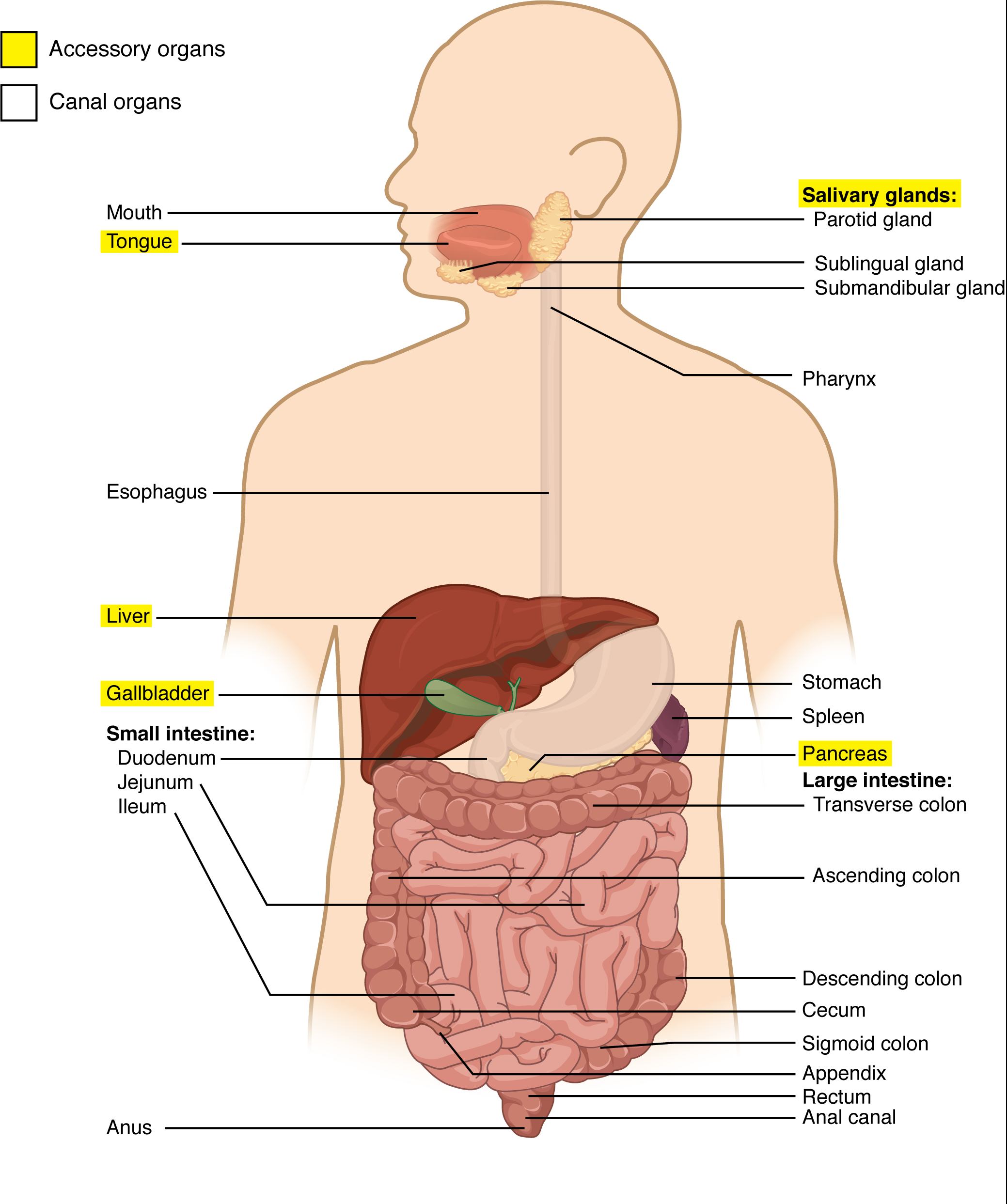
The function of the digestive system is to break down the foods you eat, release their nutrients, and absorb those nutrients into the body. Although the small intestine is the workhorse of the system, where the majority of digestion occurs, and where most of the released nutrients are absorbed into the blood or lymph, each of the digestive system organs makes a vital contribution to this process ( Figure 23.1.1 ).
As is the case with all body systems, the digestive system does not work in isolation; it functions cooperatively with the other systems of the body. Consider for example, the interrelationship between the digestive and cardiovascular systems. Arteries supply the digestive organs with oxygen and processed nutrients, and veins drain the digestive tract. These intestinal veins, constituting the hepatic portal system, are unique in that they do not return blood directly to the heart. Rather, this blood is diverted to the liver where its nutrients are off-loaded for processing before blood completes its circuit back to the heart. At the same time, the digestive system provides nutrients to the heart muscle and vascular tissue to support their functioning. The interrelationship of the digestive and endocrine systems is also critical. Hormones secreted by several endocrine glands, as well as endocrine cells of the pancreas, the stomach, and the small intestine, contribute to the control of digestion and nutrient metabolism. In turn, the digestive system provides the nutrients to fuel endocrine function. Table 23.1 gives a quick glimpse at how these other systems contribute to the functioning of the digestive system.
Digestive System Organs
The easiest way to understand the digestive system is to divide its organs into two main categories. The first group is the organs that make up the alimentary canal. Accessory digestive organs comprise the second group and are critical for orchestrating the breakdown of food and the assimilation of its nutrients into the body. Accessory digestive organs, despite their name, are critical to the function of the digestive system.
Alimentary Canal Organs
Also called the gastrointestinal (GI) tract or gut, the alimentary canal (aliment- = “to nourish”) is a one-way tube about 7.62 meters (25 feet) in length during life and closer to 10.67 meters (35 feet) in length when measured after death, once smooth muscle tone is lost. The main function of the organs of the alimentary canal is to nourish the body by digesting food and absorbing released nutrients. This tube begins at the mouth and terminates at the anus. Between those two points, the canal is modified as the pharynx, esophagus, stomach, and small and large intestines to fit the functional needs of the body. Both the mouth and anus are open to the external environment; thus, food and wastes within the alimentary canal are technically considered to be outside the body. Only through the process of absorption do the nutrients in food enter into and nourish the body’s “inner space.”
Accessory Structures
Each accessory digestive organ aids in the breakdown of food ( Figure 23.1.2 ). Within the mouth, the teeth and tongue begin mechanical digestion, whereas the salivary glands begin chemical digestion. Once food products enter the small intestine, the gallbladder, liver, and pancreas release secretions—such as bile and enzymes—essential for digestion to continue. Together, these are called accessory organs because they sprout from the lining cells of the developing gut (mucosa) and augment its function; indeed, you could not live without their vital contributions, and many significant diseases result from their malfunction. Even after development is complete, they maintain a connection to the gut by way of ducts.
Histology of the Alimentary Canal
Throughout its length, the alimentary tract is composed of the same four tissue layers; the details of their structural arrangements vary to fit their specific functions. Starting from the lumen and moving outwards, these layers are the mucosa, submucosa, muscularis, and serosa, which is continuous with the mesentery (see Figure 23.1.2 ).
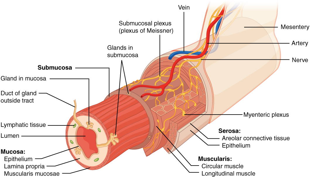
The mucosa is referred to as a mucous membrane, because mucus production is a characteristic feature of gut epithelium. The membrane consists of epithelium, which is in direct contact with ingested food, and the lamina propria, a layer of connective tissue analogous to the dermis. In addition, the mucosa has a thin, smooth muscle layer, called the muscularis mucosa (not to be confused with the muscularis layer, described below).
Epithelium —In the mouth, pharynx, esophagus, and anal canal, the epithelium is primarily a non-keratinized, stratified squamous epithelium. In the stomach and intestines, it is a simple columnar epithelium. Notice that the epithelium is in direct contact with the lumen, the space inside the alimentary canal. Interspersed among its epithelial cells are goblet cells, which secrete mucus and fluid into the lumen, and enteroendocrine cells, which secrete hormones into the interstitial spaces between cells. Epithelial cells have a very brief lifespan, averaging from only a couple of days (in the mouth) to about a week (in the gut). This process of rapid renewal helps preserve the health of the alimentary canal, despite the wear and tear resulting from continued contact with foodstuffs.
Lamina propria —In addition to loose connective tissue, the lamina propria contains numerous blood and lymphatic vessels that transport nutrients absorbed through the alimentary canal to other parts of the body. The lamina propria also serves an immune function by housing clusters of lymphocytes, making up the mucosa-associated lymphoid tissue (MALT). These lymphocyte clusters are particularly substantial in the distal ileum where they are known as Peyer’s patches. When you consider that the alimentary canal is exposed to foodborne bacteria and other foreign matter, it is not hard to appreciate why the immune system has evolved a means of defending against the pathogens encountered within it.
Muscularis mucosa —This thin layer of smooth muscle is in a constant state of tension, pulling the mucosa of the stomach and small intestine into undulating folds. These folds dramatically increase the surface area available for digestion and absorption.
As its name implies, the submucosa lies immediately beneath the mucosa. A broad layer of dense connective tissue, it connects the overlying mucosa to the underlying muscularis. It includes blood and lymphatic vessels (which transport absorbed nutrients), and a scattering of submucosal glands that release digestive secretions. Additionally, it serves as a conduit for a dense branching network of nerves, the submucosal plexus, which functions as described below.
The third layer of the alimentary canal is the muscalaris (also called the muscularis externa). The muscularis in the small intestine is made up of a double layer of smooth muscle: an inner circular layer and an outer longitudinal layer. The contractions of these layers promote mechanical digestion, expose more of the food to digestive chemicals, and move the food along the canal. In the most proximal and distal regions of the alimentary canal, including the mouth, pharynx, anterior part of the esophagus, and external anal sphincter, the muscularis is made up of skeletal muscle, which gives you voluntary control over swallowing and defecation. The basic two-layer structure found in the small intestine is modified in the organs proximal and distal to it. The stomach is equipped for its churning function by the addition of a third layer, the oblique muscle. While the colon has two layers like the small intestine, its longitudinal layer is segregated into three narrow parallel bands, the tenia coli, which make it look like a series of pouches rather than a simple tube.
The serosa is the portion of the alimentary canal superficial to the muscularis. Present only in the region of the alimentary canal within the abdominal cavity, it consists of a layer of visceral peritoneum overlying a layer of loose connective tissue. Instead of serosa, the mouth, pharynx, and esophagus have a dense sheath of collagen fibers called the adventitia. These tissues serve to hold the alimentary canal in place near the ventral surface of the vertebral column.
Nerve Supply
As soon as food enters the mouth, it is detected by receptors that send impulses along the sensory neurons of cranial nerves. Without these nerves, not only would your food be without taste, but you would also be unable to feel either the food or the structures of your mouth, and you would be unable to avoid biting yourself as you chew, an action enabled by the motor branches of cranial nerves.
Intrinsic innervation of much of the alimentary canal is provided by the enteric nervous system, which runs from the esophagus to the anus, and contains approximately 100 million motor, sensory, and interneurons (unique to this system compared to all other parts of the peripheral nervous system). These enteric neurons are grouped into two plexuses. The myenteric plexus (plexus of Auerbach) lies in the muscularis layer of the alimentary canal and is responsible for motility , especially the rhythm and force of the contractions of the muscularis. The submucosal plexus (plexus of Meissner) lies in the submucosal layer and is responsible for regulating digestive secretions and reacting to the presence of food (see Figure 23.1.2 ).
Extrinsic innervations of the alimentary canal are provided by the autonomic nervous system, which includes both sympathetic and parasympathetic nerves. In general, sympathetic activation (the fight-or-flight response) restricts the activity of enteric neurons, thereby decreasing GI secretion and motility. In contrast, parasympathetic activation (the rest-and-digest response) increases GI secretion and motility by stimulating neurons of the enteric nervous system.
Blood Supply
The blood vessels serving the digestive system have two functions. They transport the protein and carbohydrate nutrients absorbed by mucosal cells after food is digested in the lumen. Lipids are absorbed via lacteals, tiny structures of the lymphatic system. The blood vessels’ second function is to supply the organs of the alimentary canal with the nutrients and oxygen needed to drive their cellular processes.
Specifically, the more anterior parts of the alimentary canal are supplied with blood by arteries branching off the aortic arch and thoracic aorta. Below this point, the alimentary canal is supplied with blood by arteries branching from the abdominal aorta. The celiac trunk services the liver, stomach, and duodenum, whereas the superior and inferior mesenteric arteries supply blood to the remaining small and large intestines.
The veins that collect nutrient-rich blood from the small intestine (where most absorption occurs) empty into the hepatic portal system. This venous network takes the blood into the liver where the nutrients are either processed or stored for later use. Only then does the blood drained from the alimentary canal viscera circulate back to the heart. To appreciate just how demanding the digestive process is on the cardiovascular system, consider that while you are “resting and digesting,” about one-fourth of the blood pumped with each heartbeat enters arteries serving the intestines.
The Peritoneum
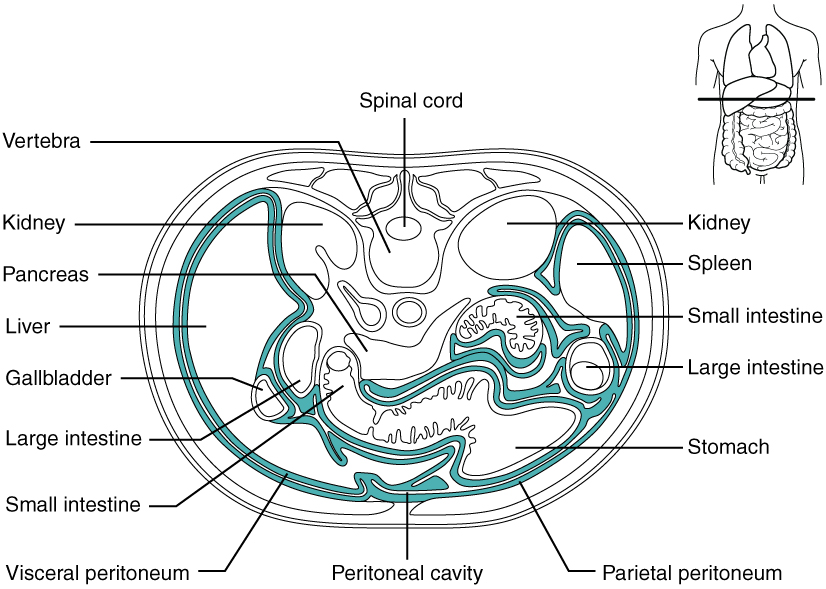
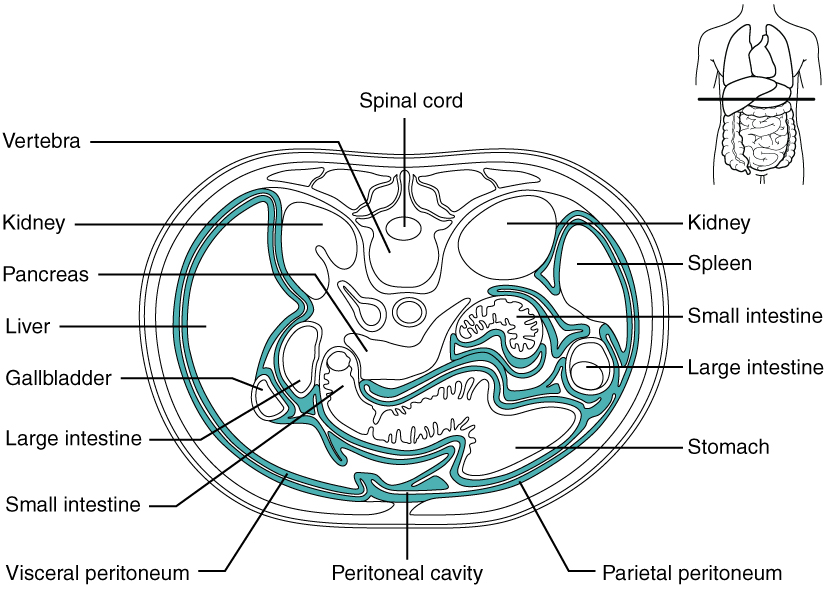
The digestive organs within the abdominal cavity are held in place by the peritoneum, a broad serous membranous sac made up of squamous epithelial tissue surrounded by connective tissue. It is composed of two different regions: the parietal peritoneum, which lines the abdominal wall, and the visceral peritoneum, which envelopes the abdominal organs ( Figure 23.1.3 ). The peritoneal cavity is the space bounded by the visceral and parietal peritoneal surfaces. A few milliliters of watery fluid act as a lubricant to minimize friction between the serosal surfaces of the peritoneum.
Inflammation of the peritoneum is called peritonitis. Chemical peritonitis can develop any time the wall of the alimentary canal is breached, allowing the contents of the lumen entry into the peritoneal cavity. For example, when an ulcer perforates the stomach wall, gastric juices spill into the peritoneal cavity. Hemorrhagic peritonitis occurs after a ruptured tubal pregnancy or traumatic injury to the liver or spleen fills the peritoneal cavity with blood. Even more severe peritonitis is associated with bacterial infections seen with appendicitis, colonic diverticulitis, and pelvic inflammatory disease (infection of uterine tubes, usually by sexually transmitted bacteria). Peritonitis is life threatening and often results in emergency surgery to correct the underlying problem and intensive antibiotic therapy. When your great grandparents and even your parents were young, the mortality from peritonitis was high. Aggressive surgery, improvements in anesthesia safety, the advance of critical care expertise, and antibiotics have greatly improved the mortality rate from this condition. Even so, the mortality rate still ranges from 30 to 40 percent.
The visceral peritoneum includes multiple large folds that envelope various abdominal organs, holding them to the dorsal surface of the body wall. Within these folds are blood vessels, lymphatic vessels, and nerves that innervate the organs with which they are in contact, supplying their adjacent organs. The five major peritoneal folds are described in Table 23.2 . An important one of these folds is the mesentery which attaches the small intestine to the body wall allowing for blood vessels, nerves, and lymphatic vessels to have a secure structure to travel through on their way to and from the small intestine. The mesocolon is the portion of the mesentery serving the colon and is considered part of the larger mesentery organ. Note that during fetal development, certain digestive structures, including the first portion of the small intestine (called the duodenum), the pancreas, and portions of the large intestine (the ascending and descending colon, and the rectum) remain completely or partially posterior to the peritoneum. Thus, the location of these organs is described as retroperitoneal .
External Website

By clicking on this link you can watch a short video of what happens to the food you eat, as it passes from your mouth to your intestine. Along the way, note how the food changes consistency and form. How does this change in consistency facilitate your gaining nutrients from food?
Chapter Review
The digestive system includes the organs of the alimentary canal and accessory structures. The alimentary canal forms a continuous tube that is open to the outside environment at both ends. The organs of the alimentary canal are the mouth, pharynx, esophagus, stomach, small intestine, and large intestine. The accessory digestive structures include the teeth, tongue, salivary glands, liver, pancreas, and gallbladder. The wall of the alimentary canal is composed of four basic tissue layers: mucosa, submucosa, muscularis, and serosa. The enteric nervous system provides intrinsic innervation, and the autonomic nervous system provides extrinsic innervation.
Interactive Link Questions
By clicking on this link , you can watch a short video of what happens to the food you eat as it passes from your mouth to your intestine. Along the way, note how the food changes consistency and form. How does this change in consistency facilitate your gaining nutrients from food?
Answers may vary.
Review Questions
Critical thinking questions.
1. Explain how the enteric nervous system supports the digestive system. What might occur that could result in the autonomic nervous system having a negative impact on digestion?
2. What layer of the alimentary canal tissue is capable of helping to protect the body against disease, and through what mechanism?
Answers for Critical Thinking Questions
- The enteric nervous system helps regulate alimentary canal motility and the secretion of digestive juices, thus facilitating digestion. If a person becomes overly anxious, sympathetic innervation of the alimentary canal is stimulated, which can result in a slowing of digestive activity.
- The lamina propria of the mucosa contains lymphoid tissue that makes up the MALT and responds to pathogens encountered in the alimentary canal.
This work, Anatomy & Physiology, is adapted from Anatomy & Physiology by OpenStax , licensed under CC BY . This edition, with revised content and artwork, is licensed under CC BY-SA except where otherwise noted.
Images, from Anatomy & Physiology by OpenStax , are licensed under CC BY except where otherwise noted.
Access the original for free at https://openstax.org/books/anatomy-and-physiology/pages/1-introduction .
Anatomy & Physiology Copyright © 2019 by Lindsay M. Biga, Staci Bronson, Sierra Dawson, Amy Harwell, Robin Hopkins, Joel Kaufmann, Mike LeMaster, Philip Matern, Katie Morrison-Graham, Kristen Oja, Devon Quick, Jon Runyeon, OSU OERU, and OpenStax is licensed under a Creative Commons Attribution-ShareAlike 4.0 International License , except where otherwise noted.

The Digestive System and Its Functions Essay
One of the most significant components of human life is digestion, because namely during this process, the necessary proteins, fats, carbohydrates, vitamins, minerals, and other useful ingredients enter the body. That is why the proper functioning of the human digestive system serves as the basis for full-fledged life support during the main processes in the digestive tract. Moreover, the digestive system is also responsible for the water-electrolytic balance, regulating the rate of fluid intake from food. The functions of the gastrointestinal tract can be summarized as follows (Hoffman 9-14):
- Motor function. Due to the middle (muscle) membrane of the digestive tract, muscle contraction-relaxation, food taking is carried out, following chewing, swallowing, mixing, and moving food along the digestive canal.
- Secretory function is carried out due to the digestive juices, that are produced by the glandular cells located in the mucous membrane (inner) of the canal. These secrets contain enzymes (reaction accelerators) that carry out the chemical processing of food (hydrolysis of food substances).
- Excretory function provides the secretion of metabolic products by the digestive glands in the gastrointestinal tract.
- Absorption function ‑ the process of assimilation of nutrients through the wall of the gastrointestinal tract into the blood and lymph.
The gastrointestinal tract is a convoluted tube that begins with the mouth and ends with the anus. The digestive system includes the following: the oral cavity with organs located in it and the adjacent large salivary glands; pharynx; esophagus; stomach; small and large intestine; liver; pancreas (Rogers 15).
The oral cavity, pharynx, and esophagus located in the area of the human head, neck, and chest cavity have a relatively straight direction. In the oral cavity, food enters the throat, where there is a cross of digestive and respiratory tracts. Then the esophagus comes, through which food mixed with saliva enters the stomach. In the oral cavity, the primary processing of food occurs, which consists of its mechanical grinding with the help of the tongue and teeth and turning into a food lump.
The salivary glands secrete saliva, the enzymes of which start the breakdown of carbohydrates in food (Smith and Morton 29). Then, through the throat and esophagus, food enters the stomach, where it is digested under the influence of gastric juice.
The stomach is a thick-walled muscle sac located under the diaphragm in the left half of the abdominal cavity. By reducing the walls of the stomach, its contents are mixed. Many glands concentrated in the mucous wall of the stomach secrete gastric juice containing enzymes and hydrochloric acid. After this, partially digested food enters the anterior part of the small intestine ‑ the duodenum.
The small intestine consists of the duodenum, jejunum, and ileum. In the duodenum, food is exposed to the action of pancreatic juice, bile, and also the juice of the glands located in its wall. In the jejunum and ileum, the final digestion of food and absorption of nutrients into the blood occurs. Undigested residues enter the colon. Here they are accumulated and are subject to removal from the body in the form of feces. The initial part of the colon is called the blind, and the appendix is following it.
Digestive glands include salivary glands, microscopic glands of the stomach and intestines, pancreas, and liver. The liver is the largest gland in the human body. It is located on the right under the diaphragm (Rogers 42). Bile is produced in the liver, which flows through the ducts into the gall bladder, where it accumulates and enters the intestine as needed. The liver retains toxic substances and protects the body from poisoning. The pancreas also belongs to the digestive glands that secrete juices and turn complex nutrients into simpler and more soluble in water. It is located between the stomach and the duodenum. Pancreatic juice contains enzymes that break down proteins, fats, and carbohydrates; 1–1.5 liters of pancreatic juice is secreted per day (Hoffman 30).
The correct sequential operation of the elements of the digestive system in time and space is ensured by regular processes of various levels. Enzymatic activity is characteristic of each section of the digestive tract and is maximum at a certain pH value of the medium. For example, in the stomach, the digestive process is carried out in an acidic environment.
Acidic content passing into the duodenum is neutralized, and intestinal digestion occurs in a neutral and slightly alkaline environment created by secrets secreted into the intestine ‑ bile, pancreatic juices, and intestinal secretions, which inactivate gastric enzymes (Smith and Morton 24). Intestinal digestion occurs in a neutral and slightly alkaline environment, first in the type of abdominal and then parietal digestion, ending with the absorption of hydrolysis products ‑ nutrients.
The degradation of nutrients by the type of cavity and parietal digestion is carried out by hydrolytic enzymes, each of which has specificity expressed to one degree or another. A set of enzymes in the secretions of the digestive glands has specific and individual characteristics, adapted to the digestion of the food that is characteristic of this region, and those nutrients that prevail in the diet.
Each digestion department has its internal environment, which serves as the basis for the functions assigned to it. The organs of the gastrointestinal tract, together with the auxiliary glands, gradually break down each component of the food, separating what the body needs and sending the rest of the absorbed food to waste. If at any of these stages a malfunction occurs, the organs and systems do not receive enough energy resources and, therefore, cannot fully perform their functions, causing an imbalance of the whole organism. Violations of the normal functioning of the digestive system can lead to the development of several diseases.
Works Cited
Hoffman, Gretchen. Digestive System . Benchmark Books, 2008.
Rogers, Kara. The Digestive System . Rosen Education Service, 2010.
Smith, Margaret E., and Dion G. Morton. The Digestive System: Systems of the Body Series . Churchill Livingstone, 2011.
- Chicago (A-D)
- Chicago (N-B)
IvyPanda. (2021, June 27). The Digestive System and Its Functions. https://ivypanda.com/essays/the-digestive-system-and-its-functions/
"The Digestive System and Its Functions." IvyPanda , 27 June 2021, ivypanda.com/essays/the-digestive-system-and-its-functions/.
IvyPanda . (2021) 'The Digestive System and Its Functions'. 27 June.
IvyPanda . 2021. "The Digestive System and Its Functions." June 27, 2021. https://ivypanda.com/essays/the-digestive-system-and-its-functions/.
1. IvyPanda . "The Digestive System and Its Functions." June 27, 2021. https://ivypanda.com/essays/the-digestive-system-and-its-functions/.
Bibliography
IvyPanda . "The Digestive System and Its Functions." June 27, 2021. https://ivypanda.com/essays/the-digestive-system-and-its-functions/.
- The Anatomy of the Pancreas
- Digestion of a Cheeseburger
- Digestive Journey of Cheeseburger
- Esophagus Anatomy and Physiology
- Development of the Chimpanzee Pancreas
- Human Digestion
- The Digestive System in the Human Body
- The Digestive System Analysis
- The Digestive System and Peptic Ulcers in Nursing
- "Salivary Gland Involvement..." Article by Liu et al.
- Microbial Biotechnology and Pharmaceutical Impact
- Bloodborne Infections: Human Immunodeficiency Virus
- Viruses: Are They Living or Non-Living?
- Animal, Plant, and Bacterial Cells' Cycles
- Eukaryotic and Prokaryotic Cells: Key Differences

- school Campus Bookshelves
- menu_book Bookshelves
- perm_media Learning Objects
- login Login
- how_to_reg Request Instructor Account
- hub Instructor Commons
- Download Page (PDF)
- Download Full Book (PDF)
- Periodic Table
- Physics Constants
- Scientific Calculator
- Reference & Cite
- Tools expand_more
- Readability
selected template will load here
This action is not available.

21.3: Digestive System Processes and Regulation
- Last updated
- Save as PDF
- Page ID 22410

- Whitney Menefee, Julie Jenks, Chiara Mazzasette, & Kim-Leiloni Nguyen
- Reedley College, Butte College, Pasadena City College, & Mt. San Antonio College via ASCCC Open Educational Resources Initiative
By the end of the section, you will be able to:
- Discuss seven fundamental activities of the digestive system, giving an example of each
- Describes the functions of each digestive organs
- Describe the difference between mechanical digestion and chemical digestion
- Describe the difference between peristalsis and segmentation
The digestive system uses mechanical and chemical activities to break food down into absorbable substances during its journey through the digestive system. Table \(\PageIndex{1}\) provides an overview of the basic functions of the digestive organs.
Digestive Processes
The processes of digestion include seven activities: ingestion, propulsion, mechanical or physical digestion, chemical digestion, secretion, absorption, and defecation.
The first of these processes, ingestion , refers to the entry of food into the alimentary canal through the mouth. There, the food is chewed and mixed with saliva secreted by salivary glands, which contains enzymes that begin breaking down the carbohydrates in the food plus some lipid digestion via lingual lipase. Chewing increases the surface area of the food and allows an appropriately sized bolus (chunk) to be produced.
Food leaves the mouth when the tongue and pharyngeal muscles propel it into the esophagus. This act of swallowing, the last voluntary act until defecation, is an example of propulsion , which refers to the movement of food through the digestive tract. It includes both the voluntary process of swallowing and the involuntary process of peristalsis. Peristalsis consists of sequential, alternating waves of contraction and relaxation of of circular and longitudinal layers of the muscularis externa (alimentary wall smooth muscles), which act to propel food along (Figure \(\PageIndex{1}\)). These waves also play a role in mixing food with digestive juices. Peristalsis is so powerful that foods and liquids you swallow enter your stomach even if you are standing on your head.

Digestion includes both mechanical and chemical processes. Mechanical digestion is a purely physical process that does not change the chemical nature of the food. Instead, it makes the food smaller to increase both surface area and mobility. It includes mastication , or chewing, as well as tongue movements that help break food into smaller bits and mix food with saliva. Although there may be a tendency to think that mechanical digestion is limited to the first steps of the digestive process, it occurs after the food leaves the mouth, as well. The mechanical churning of food in the stomach serves to further break it apart and expose more of its surface area to digestive juices, creating an acidic “soup” called chyme . Segmentation , which occurs mainly in the small intestine, consists of localized contractions of circular muscle of the muscularis layer of the alimentary canal. These contractions isolate small sections of the intestine, moving their contents back and forth while continuously subdividing, breaking up, and mixing the contents. By moving food back and forth in the intestinal lumen, segmentation mixes food with digestive juices and facilitates absorption.
Chemical digestion is aided by secretion of enzymes. Starting in the mouth, digestive secretions break down complex food molecules into their chemical building blocks (for example, proteins into separate amino acids). These secretions vary in composition, but typically contain water, various enzymes, acids, and salts. The process is completed in the small intestine.
Food that has been broken down is of no value to the body unless it enters the bloodstream and its nutrients are put to work. This occurs through the process of absorption , which takes place primarily within the small intestine. There, most nutrients are absorbed from the lumen of the alimentary canal into the bloodstream through the epithelial cells that make up the mucosa. Lipids are absorbed into lacteals and are transported via the lymphatic vessels to the bloodstream (the subclavian veins near the heart). The details of these processes will be discussed later.
In defecation , the final step in digestion, undigested materials are removed from the body as feces.
AGING AND THE...
Digestive System: From Appetite Suppression to Constipation
Age-related changes in the digestive system begin in the mouth and can affect virtually every aspect of the digestive system. Taste buds become less sensitive, so food isn’t as appetizing as it once was. A slice of pizza is a challenge, not a treat, when you have lost teeth, your gums are diseased, and your salivary glands aren’t producing enough saliva. Swallowing can be difficult, and ingested food moves slowly through the alimentary canal because of reduced strength and tone of muscular tissue. Neurosensory feedback is also dampened, slowing the transmission of messages that stimulate the release of enzymes and hormones.
Pathologies that affect the digestive organs—such as hiatal hernia, gastritis, and peptic ulcer disease—can occur at greater frequencies as you age. Problems in the small intestine may include duodenal ulcers, maldigestion, and malabsorption. Problems in the large intestine include hemorrhoids, diverticular disease, and constipation. Conditions that affect the function of accessory organs—and their abilities to deliver pancreatic enzymes and bile to the small intestine—include jaundice, acute pancreatitis, cirrhosis, and gallstones.
In some cases, a single organ is in charge of a digestive process. For example, ingestion occurs only in the mouth and defecation from the anus. However, most digestive processes involve the interaction of several organs and occur gradually as food moves through the alimentary canal (Figure \(\PageIndex{2}\)). Figure 21.3.2 shows the digestive tract with the locations of propulsion, chemical digestion, mechanical digestion, and absorption in different organs.

While most chemical digestion occurs in the small intestine, some occurs in the mouth (carbohydrates and lipids) and stomach (proteins). Absorption, also largely carried out by the small intestine, some can occur in the mouth, stomach, and large intestine. For example, alcohol and aspirin are absorbed by the stomach and water and many ions are absorbed by the large intestine.
Regulatory Mechanisms
Neural and endocrine regulatory mechanisms work to maintain the optimal conditions in the lumen needed for digestion and absorption. These regulatory mechanisms, which stimulate digestive activity through mechanical and chemical activity, are controlled both extrinsically and intrinsically.
Neural Controls
The walls of the alimentary canal contain a variety of sensors that help regulate digestive functions. These include mechanoreceptors, chemoreceptors, and osmoreceptors, which are capable of detecting mechanical, chemical, and osmotic stimuli, respectively. For example, these receptors can sense when the presence of food has caused the stomach to expand, whether food particles have been sufficiently broken down, how much liquid is present, and the type of nutrients in the food (lipids, carbohydrates, and/or proteins). Stimulation of these receptors provokes an appropriate reflex that furthers the process of digestion. This may entail sending a message that activates the glands that secrete digestive juices into the lumen, or it may mean the stimulation of muscles within the alimentary canal, thereby activating peristalsis and segmentation that move food along the intestinal tract.
The walls of the entire alimentary canal are embedded with nerve plexuses (enteric nervous system, submucosal and myenteric plexuses) that interact with the central nervous system and other nerve plexuses—either within the same digestive organ or in different ones. These interactions prompt several types of reflexes. Extrinsic nerve plexuses orchestrate long reflexes, which involve the central and autonomic nervous systems and work in response to stimuli from outside the digestive system. Short reflexes, on the other hand, are orchestrated by intrinsic nerve plexuses within the alimentary canal wall. These two plexuses and their connections were introduced earlier as the enteric nervous system. Short reflexes regulate activities in one area of the digestive tract and may coordinate local peristaltic movements and stimulate digestive secretions. For example, the sight, smell, and taste of food initiate long reflexes that begin with a sensory neuron delivering a signal to the medulla oblongata. The response to the signal is to stimulate cells in the stomach to begin secreting digestive juices in preparation for incoming food. In contrast, food that distends the stomach initiates short reflexes that cause cells in the stomach wall to increase their secretion of digestive juices.
Hormonal Controls
A variety of hormones are involved in the digestive process. The main digestive hormone of the stomach is gastrin, which is secreted in response to the presence of food. Gastrin stimulates the secretion of gastric acid by the parietal cells of the stomach mucosa. Other GI hormones are produced and act upon the gut and its accessory organs. Hormones produced by the duodenum include secretin, which stimulates a watery secretion of bicarbonate by the pancreas; cholecystokinin (CCK), which stimulates the secretion of pancreatic enzymes and bile from the liver and release of bile from the gallbladder; and gastric inhibitory peptide, which inhibits gastric secretion and slows gastric emptying and motility. These GI hormones are secreted by specialized epithelial cells, called enteroendocrine cells, located in the mucosal epithelium of the stomach and small intestine. These hormones then enter the bloodstream, through which they can reach their target organs.
Concept Review
The digestive system ingests and digests food, absorbs released nutrients, and excretes food components that are indigestible. The six activities involved in this process are ingestion (mouth), motility (GI tract), mechanical digestion (mouth, stomach, small intestine), chemical digestion (mouth, stomach, small intestine), absorption (mouth, stomach, small and large intestines), and defecation (anus). Contractions of smooth muscles (muscularis externa) result in peristalsis to push contents along in the GI tract and segmentation to mix the content with enzymes. These processes are regulated by neural and hormonal mechanisms.
Review Questions
Q. Which of these processes occurs in the mouth?
A. ingestion
B. mechanical digestion
C. chemical digestion
D. all of the above
Q. Which of these processes occurs throughout most of the alimentary canal?
B. propulsion
C. segmentation
D. absorption
Q. Which of the following occur(s) in the mouth?
A. mechanical digestion
B. chemical digestion
C. mastication
Q. Which of these statements about the colon is false?
A. Chemical digestion occurs in the colon.
B. Absorption occurs in the colon.
C. Peristalsis occurs in the colon.
D. Diverticular disease occurs in the colon.
Critical Thinking Questions
Q. Offer a theory to explain why segmentation occurs and peristalsis slows in the small intestine.
A. The majority of digestion and absorption occurs in the small intestine. By slowing the transit of chyme, segmentation and a reduced rate of peristalsis allow time for these processes to occur.
Q. Which organ is mostly responsible for diarrhea and constipation and why?
A. The colon absorbs water. If it absorbs too much water, then the remaining contents (stool) may be hard and constipation may result. If it absorbs very little water or even secretes water, then the remaining contents will be loose and watery, resulting in diarrhea.
Contributors and Attributions
OpenStax Anatomy & Physiology (CC BY 4.0). Access for free at https://openstax.org/books/anatomy-and-physiology
- Skip to main content
- Skip to secondary menu
- Skip to primary sidebar
- Skip to footer
Study Today
Largest Compilation of Structured Essays and Exams
Human Digestive System | Essay for Children & Students
December 16, 2017 by Study Mentor Leave a Comment
The digestive system is a system of organs working together for the uptake of food, its digestion and to eliminate the indigestible waste products out of the body. It is also known as the Alimentary canal.
Table of Contents
What is digestion?
Digestion is defined as the breakdown of complex organic food materials into simpler compounds and thus process, releasing energy required by the body.
The complex food substances are carbohydrates, fats, proteins, vitamins, minerals etc. Digestion involves two processes: Mechanical digestion in which food is broken down by the peristaltic movement of the organs involved in digestion and chemical digestion in which digestive juices act on the complex food materials to convert it into simpler compounds.
Essential compounds of diet
- Water : Water is a very important compound which can be obtained from all food and drinks and is also released in the body due to oxidation of food. The optimum requirement of water for an adult is 2,000ml.
- Carbohydrates : Carbohydrates can be obtained from food sources like cereals, bread, fruits etc. They help in providing energy.
- Protein : Proteins help in growth and maintenance of the body. It also helps in formation of enzymes. Proteins can be obtained from pulses, milk, meat, eggs, cheese etc.
- Fats/Lipids : It also helps in providing energy. It can be obtained from milk, butter, ghee, oil, creams, nuts etc.
- Vitamins : Vitamins help in prevention of deficiency diseases and regulation of metabolic activities. The vitamins required by our body are A, B, B6, B12, C, D, E and K.
- Minerals : Different minerals have different functions in the human body. Some of the important minerals are-
- Sodium : Helps in osmotic balance, muscle contraction and nerve impulse conduction. It can be obtained from table salt and vegetables.
- Iron : Oxygen transport as part of haemoglobin. Can be obtained from leafy vegetables and iron supplements.
- Iodine : It helps in metabolic control of hormone thyroxine.
- Potassium : Helps in muscle contraction and nerve impulse conduction. Can be obtained from vegetables.
The human digestive system consists of the alimentary canal and associated digestive glands.
Alimentary canal
Histology. The alimentary canal is lined with muscular layers. It consists of four layers.
- Serosa: It is the outermost layer. It is formed of a single layer of cells.
- Muscularised: It is formed by two layers of cells and consists of a network of nerve cells. It controls the muscular contractions.
- Submucosa: It consists of highly vascular connective tissue. It also has another network of nerve fibres, it controls the secretion of intestinal juice.
- Mucosa: It is the innermost layer and consists of further 3 layers. It forms the digestive juices.
Parts of the alimentary canal

Buccal cavity: It is the space which is bounded to the sides by jaws, top by the palate and below by the throat. The buccal cavity consists of the tongue which is a highly muscular organ and consists of papillae. The tongue helps in mixing saliva with food and facilitates in swallowing. It also helps in telling the taste of the food.
Teeth : Teeth are embedded in both the upper and lower jaw and helps in chewing, cutting and piercing the food. There are four types of teeth present in humans – Incisors, canines, premolars and molars. Adult human being consists of 32 teeth in the permanent set.
A tooth consists of 3 regions- Crown, neck and root. The root is embedded completely in the jaw and consists of nerve endings and blood vessels. The neck is surrounded by the gum which is soft and fleshy skin. The crown is the exposes part of the tooth and is covered by a shiny material called enamel.
Pharynx : The food and air crosses the pharynx to reach the oesophagus. It has voluntary muscles which contract and help in swallowing.
Oesophagus : It is the long, muscular straight tube which connects the pharynx to the stomach. The major function of the oesophagus is to pass the food from pharynx to the stomach by peristaltic movement.
Stomach . The stomach is a J shaped muscular sac which stores the food for some time. It is also involved in mechanical churning of food, partial digestion of food by gastric juices and regulation of passage of food in the small intestine.
It has 3 parts- The cardiac, fundic and pyloric. The inner surface of the stomach consists of various folding known as gastric rugae which help in increasing the surface area for maximum storage of food. Stomach also secretes the hormone gastrin.
Small intestine: Divided into 3 major parts- duodenum, jejunum and ileum, the major function of the small intestine is absorption of food. It also consists of microscopic finger like projections known as villi which increases surface area for effective absorption. Small intestine also secretes some hormones.
Large intestine: The large intestine is divided into 3 major parts- Caecum, Colon and Rectum. Its major role is absorption of water, formation of faeces, and production of mucus for the lubrication of mucosa.
Anus : The function of the anus is elimination of faeces. It consists of two anal sphincters: the internal anal sphincter and the external anal sphincter.
Digestive glands
Human digestive glands include salivary glands, gastric glands, liver, pancreas and intestinal glands.
- Salivary glands: The function of salivary glands is to secrete saliva which is digestive in function. Saliva is secreted by 3 pairs of salivary glands-
- Parotid glands which lie on the sides of the face. The saliva produced by these glands is carried by Stensen’s duct.
- Submaxillary glands which lie at a certain angle of the lower jaw. It has submaxillary ducts which open under the tongue.
- Submandibular glands: These glands are present under the front part of the tongue. Ducts of Rivinus which carries the saliva produced by these glands open under the tongue.
- The Saliva secreted by the salivary glands has a pH of 7 and contains salivary amylase which is an enzyme. Saliva also contains lysozyme which is anti bacterial in function.
- Gastric glands: gastric glands are present in the stomach and is acidic having a pH of 2. There are 3 types of gastric glands present in the stomach:
- Mucous cells which secrete mucus that helps in protection of the internal wall of the stomach from the gastric acid.
- Peptic or chief cells which secrete pepsinogen which is the precursor of enzyme pepsin.
- Oxyntic cells which secrete hydrochloric acid.
Liver : It is the largest gland of the body consisting of hepatocytes, bile canaliculi and hepatic sinusoids. The liver weighs 1.6kg. There are two lobes in the liver. Right and left lobe.
The liver forms bile which is stored in concentrated form in the gall bladder. Liver also has a function in detoxification of poison or toxic substances in the body.
Pancreas : The pancreas is a gland which is both endocrine and exocrine in function. The endocrine part secretes hormones namely, insulin, glucagon and somatostatin.
The exocrine part secretes pancreatic juice which consists of proenzyme trypsinogen, chymotrypsinogen and procarboxypeptidase. There are other enzymes also such as pancreatic lipase, nucleases and pancreatic amylase.
Intestinal juice : the wall of villi present in the small intestine contains small, microscopic glands, Brunner’s glands and Crypts if Lieberkühn.
The both secrete enzymes, mucus and alkaline watery fluid. The mixture of all these secretions is known as the intestinal juice or succus entericus.
Digestion of food

The organs churn the food by mass peristaltic movement and the digestive glands pour their secretions to facilitate the process of digestion. The processes of digestion in various organs are as follows:
Mouth and buccal cavity: Digestion of starch starts in the mouth where starch is converted into maltose by the action of enzyme, salivary amylase. The saliva also contains various electrolytes such as sodium, potassium, chlorine etc. 30% of starch is hydrolysed here and the food is converted into a bolus which is further passed down to the Oesophagus.
Oesophagus : It does not contain any digestive gland so it does not aid in digestion. It only helps in passage of food from the buccal cavity to the stomach.
Stomach . The stomach consists of Gastric glands which secrete gastric juice. The gastric juice gets mixed with the bolus by the churning movements of the stomach. HCl helps in conversion of proenzyme pepsinogen into active form, pepsin. Pepsin helps in hydrolysis of proteins into peptides. Digestion of casein present in milk also takes place in the stomach by the action of enzyme rennin.
Small intestine : The small intestine plays a major role in both digestion and absorption of food. The pancreatic juice and bile from the gall bladder are released into the small intestine. Bile juice helps in emulsification of fats. It also coats each small fat droplet to avoid their merging together. The Pancreatic amylase hydrolyses starch and glycogen into maltose and dextrin’s.
Enzyme trypsinogen gets activated into trypsin by enterokinase. Trypsin converts proteins to peptides. It also converts chymotrypsinogen to chymotrypsin. Trypsin also helps in activation of procarboxypeptidase into carboxypeptidases which further converts peptides into amino acids.
After digestion, food is absorption of nutrition from food and its passage into the blood and the lymph. In mouth, very little absorption takes place. Absorption of few drugs takes place in the mouth into the buccal mucosa. Absorption of simple sugars, water, and alcohol takes place in the stomach.
Major absorption takes place in the small intestine. The final products after digestion such as glucose, fatty acids, amino acids etc. get absorbed into the blood lymph. The large intestine is also not much involved in absorption. It only absorbs some minerals and water.
Reader Interactions
Leave a reply cancel reply.
Your email address will not be published. Required fields are marked *
Top Trending Essays in March 2021
- Essay on Pollution
- Essay on my School
- Summer Season
- My favourite teacher
- World heritage day quotes
- my family speech
- importance of trees essay
- autobiography of a pen
- honesty is the best policy essay
- essay on building a great india
- my favourite book essay
- essay on caa
- my favourite player
- autobiography of a river
- farewell speech for class 10 by class 9
- essay my favourite teacher 200 words
- internet influence on kids essay
- my favourite cartoon character
Brilliantly
Content & links.
Verified by Sur.ly
Essay for Students
- Essay for Class 1 to 5 Students
Scholarships for Students
- Class 1 Students Scholarship
- Class 2 Students Scholarship
- Class 3 Students Scholarship
- Class 4 Students Scholarship
- Class 5 students Scholarship
- Class 6 Students Scholarship
- Class 7 students Scholarship
- Class 8 Students Scholarship
- Class 9 Students Scholarship
- Class 10 Students Scholarship
- Class 11 Students Scholarship
- Class 12 Students Scholarship
STAY CONNECTED
- About Study Today
- Privacy Policy
- Terms & Conditions
Scholarships
- Apj Abdul Kalam Scholarship
- Ashirwad Scholarship
- Bihar Scholarship
- Canara Bank Scholarship
- Colgate Scholarship
- Dr Ambedkar Scholarship
- E District Scholarship
- Epass Karnataka Scholarship
- Fair And Lovely Scholarship
- Floridas John Mckay Scholarship
- Inspire Scholarship
- Jio Scholarship
- Karnataka Minority Scholarship
- Lic Scholarship
- Maulana Azad Scholarship
- Medhavi Scholarship
- Minority Scholarship
- Moma Scholarship
- Mp Scholarship
- Muslim Minority Scholarship
- Nsp Scholarship
- Oasis Scholarship
- Obc Scholarship
- Odisha Scholarship
- Pfms Scholarship
- Post Matric Scholarship
- Pre Matric Scholarship
- Prerana Scholarship
- Prime Minister Scholarship
- Rajasthan Scholarship
- Santoor Scholarship
- Sitaram Jindal Scholarship
- Ssp Scholarship
- Swami Vivekananda Scholarship
- Ts Epass Scholarship
- Up Scholarship
- Vidhyasaarathi Scholarship
- Wbmdfc Scholarship
- West Bengal Minority Scholarship
- Click Here Now!!
Mobile Number
Have you Burn Crackers this Diwali ? Yes No
Want to create or adapt books like this? Learn more about how Pressbooks supports open publishing practices.
Chapter 12 Digestive System
12.1 Overview of the Digestive System
By the end of this section, you will be able to:
Identify the organs of the alimentary canal from proximal to distal, and briefly state their function
Identify the accessory digestive organs and briefly state their function
Describe the four fundamental tissue layers of the alimentary canal
Contrast the contributions of the enteric and autonomic nervous systems to digestive system functioning
Explain how the peritoneum anchors the digestive organs
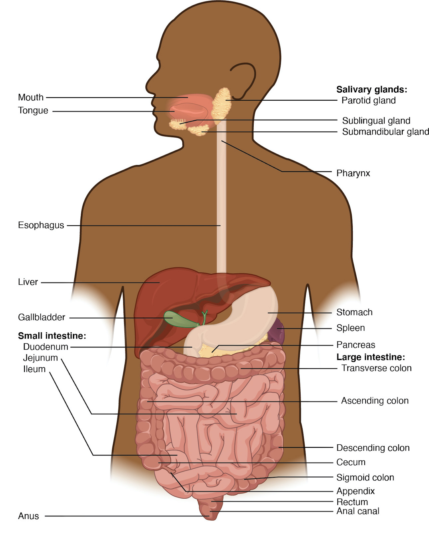
The function of the digestive system is to break down the foods you eat, release their nutrients, and absorb those nutrients into the body. Although the small intestine is the workhorse of the system, where the majority of digestion occurs, and where most of the released nutrients are absorbed into the blood or lymph, each of the digestive system organs makes a vital contribution to this process (Figure 12.2 ).
As is the case with all body systems, the digestive system does not work in isolation; it functions cooperatively with the other systems of the body. Consider for example, the interrelationship between the digestive and cardiovascular systems. Arteries supply the digestive organs with oxygen and processed nutrients, and veins drain the digestive tract. These intestinal veins, constituting the hepatic portal system, are unique; they do not return blood directly to the heart. Rather, this blood is diverted to the liver where its nutrients are off-loaded for processing before blood completes its circuit back to the heart. At the same time, the digestive system provides nutrients to the heart muscle and vascular tissue to support their functioning. The interrelationship of the digestive and endocrine systems is also critical. Hormones secreted by several endocrine glands, as well as endocrine cells of the pancreas, the stomach, and the small intestine, contribute to the control of digestion and nutrient metabolism. In turn, the digestive system provides the nutrients to fuel endocrine function. Table 12.1 gives a quick glimpse at how these other systems contribute to the functioning of the digestive system.
The easiest way to understand the digestive system is to divide its organs into two main categories. The first group is the organs that make up the alimentary canal. Accessory digestive organs comprise the second group and are critical for orchestrating the breakdown of food and the assimilation of its nutrients into the body. Accessory digestive organs, despite their name, are critical to the function of the digestive system.
Alimentary Canal Organs
Also called the gastrointestinal (GI) tract or gut, the alimentary canal (aliment- = “to nourish”) is a one-way tube about 7.62 meters (25 feet) in length during life and closer to 10.67 meters (35 feet) in length when measured after death, once smooth muscle tone is lost. The main function of the organs of the alimentary canal is to nourish the body. This tube begins at the mouth and terminates at the anus. Between those two points, the canal is modified as the pharynx, esophagus, stomach, and small and large intestines to fit the functional needs of the body. Both the mouth and anus are open to the external environment; thus, food and wastes within the alimentary canal are technically considered to be outside the body. Only through the process of absorption do the nutrients in food enter into and nourish the body’s “inner space.”
Accessory Structures
Each accessory digestive organ aids in the breakdown of food (Figure 12.2 ). Within the mouth, the teeth and tongue begin mechanical digestion, whereas the salivary glands begin chemical digestion. Once food products enter the small intestine, the gallbladder, liver, and pancreas release secretions — such as bile and enzymes — essential for digestion to continue. Together, these are called accessory organs because they sprout from the lining cells of the developing gut (mucosa) and augment its function; indeed, you could not live without their vital contributions, and many significant diseases result from their malfunction. Even after development is complete, they maintain a connection to the gut by way of ducts.
Histology of the Alimentary Canal
Throughout its length, the alimentary tract is composed of the same four tissue layers; the details of their structural arrangements vary to fit their specific functions. Starting from the lumen and moving outwards, these layers are the mucosa, submucosa, muscularis, and serosa, which is continuous with the mesentery (see Figure 12.3 ).
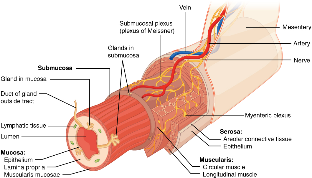
The mucosa is referred to as a mucous membrane, because mucus production is a characteristic feature of gut epithelium. The membrane consists of epithelium, which is in direct contact with ingested food, and the lamina propria, a layer of connective tissue. In addition, the mucosa has a thin, smooth muscle layer, called the muscularis mucosae (not to be confused with the muscularis layer, described below).
Epithelium — In the mouth, pharynx, esophagus, and anal canal, the epithelium is primarily a non-keratinized, stratified squamous epithelium. In the stomach and intestines, it is a simple columnar epithelium. The epithelium is in direct contact with the lumen, the space inside the alimentary canal. Interspersed among its epithelial cells are goblet cells, which secrete mucus and fluid into the lumen, and enteroendocrine cells, which secrete hormones into the interstitial spaces between cells. Epithelial cells have a very brief lifespan, averaging from only a couple of days (in the mouth) to about a week (in the gut). This process of rapid renewal helps preserve the health of the alimentary canal, despite the wear and tear resulting from continued contact with foodstuffs.
Lamina propria — In addition to loose connective tissue, the lamina propria contains numerous blood and lymphatic vessels that transport nutrients absorbed through the alimentary canal to other parts of the body. The lamina propria also serves an immune function by housing clusters of lymphocytes, making up the mucosa-associated lymphoid tissue (MALT). These lymphocyte clusters are particularly substantial in the distal ileum where they are known as Peyer’s patches. When you consider that the alimentary canal is exposed to foodborne bacteria and other foreign matter, it is not hard to appreciate why the immune system has evolved a means of defending against the pathogens encountered within it.
Muscularis mucosae — This thin layer of smooth muscle is in a constant state of tension, pulling the mucosa of the stomach and small intestine into undulating folds. These folds dramatically increase the surface area available for digestion and absorption.
As its name implies, the submucosa lies immediately beneath the mucosa. A broad layer of dense connective tissue, it connects the overlying mucosa to the underlying muscularis. It includes blood and lymphatic vessels (which transport absorbed nutrients), and a scattering of submucosal glands that release digestive secretions. Additionally, it serves as a conduit for a dense branching network of nerves, the submucosal plexus, which functions as described below.
The third layer of the alimentary canal is the muscularis (also called the muscularis externa). The muscularis in the small intestine is made up of a double layer of smooth muscle: an inner circular layer and an outer longitudinal layer. The contractions of these layers promote mechanical digestion, expose more of the food to digestive chemicals, and move the food along the canal. In the most proximal and distal regions of the alimentary canal, including the mouth, pharynx, anterior part of the esophagus, and external anal sphincter, the muscularis is made up of skeletal muscle, which gives you voluntary control over swallowing and defecation. The basic two-layer structure found in the small intestine is modified in the organs proximal and distal to it. The stomach is equipped for its churning function by the addition of a third layer, the oblique muscle. While the colon has two layers like the small intestine, its longitudinal layer is segregated into three narrow parallel bands, the tenia coli, which make it look like a series of pouches rather than a simple tube.
The serosa is the portion of the alimentary canal superficial to the muscularis. Present only in the region of the alimentary canal within the abdominal cavity, it consists of a layer of visceral peritoneum overlying a layer of loose connective tissue. Instead of serosa, the mouth, pharynx, and esophagus have a dense sheath of collagen fibers called the adventitia. These tissues serve to hold the alimentary canal in place near the ventral surface of the vertebral column.
Nerve Supply
As soon as food enters the mouth, it is detected by receptors that send impulses along the sensory neurons of cranial nerves. Without these nerves, not only would your food be without taste, but you would also be unable to feel either the food or the structures of your mouth, and you would be unable to avoid biting yourself as you chew, an action enabled by the motor branches of cranial nerves. Intrinsic innervation of much of the alimentary canal is provided by the enteric nervous system, which runs from the esophagus to the anus, and contains approximately 100 million motor, sensory, and interneurons (unique to this system compared to all other parts of the peripheral nervous system). These enteric neurons are grouped into two plexuses. The myenteric plexus (plexus of Auerbach) lies in the muscularis layer of the alimentary canal and is responsible for motility , especially the rhythm and force of the contractions of the muscularis. The submucosal plexus (plexus of Meissner) lies in the submucosal layer and is responsible for regulating digestive secretions and reacting to the presence of food (see 12.3 ).
Extrinsic innervations of the alimentary canal are provided by the autonomic nervous system, which includes both sympathetic and parasympathetic nerves. In general, sympathetic activation (the fight-or-flight response) restricts the activity of enteric neurons, thereby decreasing GI secretion and motility. In contrast, parasympathetic activation (the rest-and-digest response) increases GI secretion and motility by stimulating neurons of the enteric nervous system.
Blood Supply
The blood vessels serving the digestive system have two functions. They transport the protein and carbohydrate nutrients absorbed by mucosal cells after food is digested in the lumen. Lipids are absorbed via lacteals, tiny structures of the lymphatic system. The blood vessels’ second function is to supply the organs of the alimentary canal with the nutrients and oxygen needed to drive their cellular processes.
Specifically, the more anterior parts of the alimentary canal are supplied with blood by arteries branching off the aortic arch and thoracic aorta. Below this point, the alimentary canal is supplied with blood by arteries branching from the abdominal aorta. The celiac trunk services the liver, stomach, and duodenum, whereas the superior and inferior mesenteric arteries supply blood to the remaining small and large intestines.
The veins that collect nutrient-rich blood from the small intestine (where most absorption occurs) empty into the hepatic portal system. This venous network takes the blood into the liver where the nutrients are either processed or stored for later use. Only then does the blood drained from the alimentary canal viscera circulate back to the heart. To appreciate just how demanding the digestive process is on the cardiovascular system, consider that while you are “resting and digesting,” about one-fourth of the blood pumped with each heartbeat enters arteries serving the intestines.
The Peritoneum
The digestive organs within the abdominal cavity are held in place by the peritoneum, a broad serous membranous sac made up of squamous epithelial tissue surrounded by connective tissue. It is composed of two different regions: the parietal peritoneum, which lines the abdominal wall, and the visceral peritoneum, which envelopes the abdominal organs ( Figure 12.4 ). The peritoneal cavity is the space bounded by the visceral and parietal peritoneal surfaces. A few milliliters of watery fluid act as a lubricant to minimize friction between the serosal surfaces of the peritoneum.
Peritonitis
Inflammation of the peritoneum is called peritonitis. Chemical peritonitis can develop any time the wall of the alimentary canal is breached, allowing the contents of the lumen entry into the peritoneal cavity. For example, when an ulcer perforates the stomach wall, gastric juices spill into the peritoneal cavity. Hemorrhagic peritonitis occurs after a ruptured tubal pregnancy or traumatic injury to the liver or spleen fills the peritoneal cavity with blood. Even more severe peritonitis is associated with bacterial infections seen with appendicitis, colonic diverticulitis, and pelvic inflammatory disease (infection of uterine tubes, usually by sexually transmitted bacteria). Peritonitis is life threatening and often results in emergency surgery to correct the underlying problem and intensive antibiotic therapy. When your great grandparents and even your parents were young, the mortality from peritonitis was high. Aggressive surgery, improvements in anesthesia safety, the advance of critical care expertise, and antibiotics have greatly improved the mortality rate from this condition. Even so, the mortality rate still ranges from 30 to 40 percent.
The visceral peritoneum includes multiple large folds that envelope various abdominal organs, holding them to the dorsal surface of the body wall. Within these folds are blood vessels, lymphatic vessels, and nerves that innervate the organs with which they are in contact, supplying their adjacent organs.
Meet the gastrointestinal tract!
This video shows what happens to the food you eat, as it passes from your mouth to your intestine.
Along the way, note how the food changes consistency and form. How does this change in consistency facilitate your gaining nutrients from food?
Section Review
The digestive system includes the organs of the alimentary canal and accessory structures. The alimentary canal forms a continuous tube that is open to the outside environment at both ends. The organs of the alimentary canal are the mouth, pharynx, esophagus, stomach, small intestine, and large intestine. The accessory digestive structures include the teeth, tongue, salivary glands, liver, pancreas, and gallbladder. The wall of the alimentary canal is composed of four basic tissue layers: mucosa, submucosa, muscularis, and serosa. The enteric nervous system provides intrinsic innervation, and the autonomic nervous system provides extrinsic innervation.
Review Questions
Introduction to Human Biology Copyright © by Wolf T Pecher is licensed under a Creative Commons Attribution-NonCommercial-ShareAlike 4.0 International License , except where otherwise noted.
Share This Book
Home — Essay Samples — Nursing & Health — Digestive System — The Digestive Process of Food in the Alimentary Canal and Accessory Digestive Organs in the Digestive System of the Human Body
The Digestive Process of Food in The Alimentary Canal and Accessory Digestive Organs in The Digestive System of The Human Body
- Categories: Digestive System
About this sample

Words: 1362 |
Published: Dec 11, 2018
Words: 1362 | Pages: 3 | 7 min read

Cite this Essay
Let us write you an essay from scratch
- 450+ experts on 30 subjects ready to help
- Custom essay delivered in as few as 3 hours
Get high-quality help

Dr. Karlyna PhD
Verified writer
- Expert in: Nursing & Health

+ 120 experts online
By clicking “Check Writers’ Offers”, you agree to our terms of service and privacy policy . We’ll occasionally send you promo and account related email
No need to pay just yet!
Related Essays
4 pages / 1752 words
7 pages / 3233 words
2 pages / 858 words
2 pages / 1178 words
Remember! This is just a sample.
You can get your custom paper by one of our expert writers.
121 writers online
Still can’t find what you need?
Browse our vast selection of original essay samples, each expertly formatted and styled
The digestive system is a complex network of organs and tissues that work together to break down and absorb nutrients from the food we consume. This system exemplifies the interdependency and interconnectivity between structure [...]
Achlorhydria occurs when there’s an absence of hydrochloric (HCl) acids in the stomach. It’s a more severe form of a hypochlorhydria, a deficiency of stomach acids. Both conditions can impair the digestive process and lead to [...]
Ehrlich, S. D. (2013). Microbes in the gut: probiotics, prebiotics, symbionts to control inflammatory conditions. Immunity, inflammation, and disease, 1(2), 109-123.Ensminger, A. H. (2018). Foods & Nutrition Encyclopedia, 2nd [...]
The mouth chews the food using the teeth to tear, chew and grind the food. The tongue has skeletal muscles (involuntary muscles) that move the food around the mouth for efficient mechanical digestion. Salivary glands beneath and [...]
Hemostasis is the process that occurs when a blood vessel ruptures and large amounts of plasma and formed elements may escape (Bostwick and Wingerd, 2013). It can be divided into primary and secondary hemostasis. Primary [...]
In Godric, Frederick Buechner uses multiple characters who are at once medieval and modern to not only tell the uncommon tale of a flawed saint, but to depict through medieval text and setting his modern comments, [...]
Related Topics
By clicking “Send”, you agree to our Terms of service and Privacy statement . We will occasionally send you account related emails.
Where do you want us to send this sample?
By clicking “Continue”, you agree to our terms of service and privacy policy.
Be careful. This essay is not unique
This essay was donated by a student and is likely to have been used and submitted before
Download this Sample
Free samples may contain mistakes and not unique parts
Sorry, we could not paraphrase this essay. Our professional writers can rewrite it and get you a unique paper.
Please check your inbox.
We can write you a custom essay that will follow your exact instructions and meet the deadlines. Let's fix your grades together!
Get Your Personalized Essay in 3 Hours or Less!
We use cookies to personalyze your web-site experience. By continuing we’ll assume you board with our cookie policy .
- Instructions Followed To The Letter
- Deadlines Met At Every Stage
- Unique And Plagiarism Free

COMMENTS
Essay # 1. Organs of Digestive System: Digestion means simplification of complex foods. It is the process of breaking various foodstuff into simple products. The complex foods like carbohydrates, proteins and fats are converted into glucose, amino acids and fatly acids respectively by the action of digestive enzymes.
Chemical digestion is the mechanism involving processing the food bolus with the help of different substances produced by the human body such as saliva or digestive juices (Martinez, 2014). Due to chemical digestion, the organism manages to extract the nutrients that need to be absorbed. Mechanical digestion is the process that involves ...
The Stomach. The stomach is an organ of digestion located between the esophagus and the duodenum on the left upper part of the abdominal cavity. It is a muscular, elastic pear-shaped bag lying just below the diaphragm. It is made up of five layers. The innermost layer is referred to as mucosa.
human digestive system, system used in the human body for the process of digestion.The human digestive system consists primarily of the digestive tract, or the series of structures and organs through which food and liquids pass during their processing into forms that can be absorbed into the bloodstream. The system also consists of the structures through which wastes pass in the process of ...
The stomach is a rounded, hollow organ which is located just inferior to the diaphragm in the left side of the abdominal cavity. It is located between the oesophagus and duodenum, it is a rough crescent-shaped enlargement of the gastrointestinal tract. The inner layer has wrinkles known as gastric folds. The gastric folds allow the stomach to ...
It enables the organism get nutrients which are important for functioning. The most important organs of the human digestive system are mouth, salivary glands, pharynx, oesophagus, stomach, liver, pancreas, gallbladder, small intestine, large intestine and anus (Grosvenor & Smolin, 2012). Food travels through the digestive system and each organ ...
One of the vital organ systems is the digestive one and consists of the gastrointestinal tract and organs such as the tongue, salivary glands, pancreas, liver, and gallbladder (Crash Course, 2015). This organ system is responsible for processing the food that is ingested in the mouth and is located mainly in the stomach.
The digestive system is divided into two main parts: the alimentary canal (or gastrointestinal tract) and the accessory digestive organs. The alimentary canal is a continuous, coiled, hollow muscular tube that winds through the body, extending from the mouth to the anus. It includes the mouth, pharynx, esophagus, stomach, small intestine, and ...
The digestive system is a complex network of organs that work together to break down food, absorb nutrients, and eliminate waste. Learn about the anatomy, functions, and clinical aspects of the digestive system with Kenhub, a comprehensive online learning platform for anatomy and histology. Kenhub offers interactive quizzes, videos, articles, and atlas images to help you master the digestive ...
The human digestive system is responsible for the intake, breakdown, absorption and finally removal of nutrients and energy needed for the functioning of the human body. It performs these duties by allowing nutrients and energy in the form of food to enter the body, and then removing the materials needed from the food, then absorbing the ...
The Digestive System: An Introduction. The digestive system is a group of organs that work together to change the food we eat into energy our bodies can use. It's like a food processing factory inside our body. It includes the mouth, esophagus, stomach, small intestine, large intestine, rectum, and anus.
The main organs that make up your digestive system are the organs known as your gastrointestinal tract. They are: your mouth, esophagus, stomach, small intestine, large intestine and anus. Assisting your GI organs along the way are your pancreas, gallbladder and liver. Here's how these organs work together in your digestive system.
The function of the digestive system is to break down the foods you eat, release their nutrients, and absorb those nutrients into the body. Although the small intestine is the workhorse of the system, where the majority of digestion occurs, and where most of the released nutrients are absorbed into the blood or lymph, each of the digestive system organs makes a vital contribution to this ...
The breaking down of food by physical means, such as chewing. Absorption. The process by which nutrients pass through the walls of the digestive system into the blood. Excretory system. The body system that removes metabolic wastes from the body. Excretion. The process of removing wastes and excess water from the body.
That is why the proper functioning of the human digestive system serves as the basis for full-fledged life support during the main processes in the digestive tract. Moreover, the digestive system is also responsible for the water-electrolytic balance, regulating the rate of fluid intake from food. The functions of the gastrointestinal tract can ...
digestion, the breakdown of food, for example, through chewing and secretions. the absorption of nutrients, which mainly occurs in the small intestine. defecation and urination, the removal of ...
In some cases, a single organ is in charge of a digestive process. For example, ingestion occurs only in the mouth and defecation from the anus. However, most digestive processes involve the interaction of several organs and occur gradually as food moves through the alimentary canal (Figure \(\PageIndex{2}\)).
Essay on Human Digestive System. The digestive system is a system of organs working together for the uptake of food, its digestion. ... Liver: It is the largest gland of the body consisting of hepatocytes, bile canaliculi and hepatic sinusoids. The liver weighs 1.6kg. There are two lobes in the liver.
The function of the digestive system is to break down the foods you eat, release their nutrients, and absorb those nutrients into the body. Although the small intestine is the workhorse of the system, where the majority of digestion occurs, and where most of the released nutrients are absorbed into the blood or lymph, each of the digestive system organs makes a vital contribution to this ...
The Damages of Eating Disorders. 2 pages / 820 words. The digestive system includes mouth, salivary glands, liver, gallbladder, appendix esophagus, stomach, pancreas, large intestine and small intestine. The mouth starts the digestive system by the mouth breaking up food. The throat swallows the food.
Human digestive system. The human digestive system is a complex series of organs and processes that break down food into nutrients that can be absorbed and used by the body. In this essay, we will explore the anatomy and physiology of the digestive system and its role in maintaining overall health.
The major digestive organ the small intestine, an organ that is actually longer than the so called large intestine. The mall intestine is a muscular tube coming from the pyloric sphincter to the ileocecal valve, Consisting of 3 subdivisions the duodenum, jejunum, and ileum, contributes five percent, almost forty percent, to almost sixty percent of the length of the small intestine.
Anus. Study with Quizlet and memorize flashcards containing terms like Start with your mouth and ending at your anus, list in order the alimentary canal structures that food passes through during digestion., State the four layers found in these tube organs from superficial to deep., State the two types of motion used to move the food. and more.